Home

Ruisseau buffet Égoïsme cornea slit lamp Accumulation Paiement Carrière

Corneal Pachymetry and Endothelial Microscopy by Slit-Lamp | IntechOpen

How to Use a Slit Lamp - American Academy of Ophthalmology

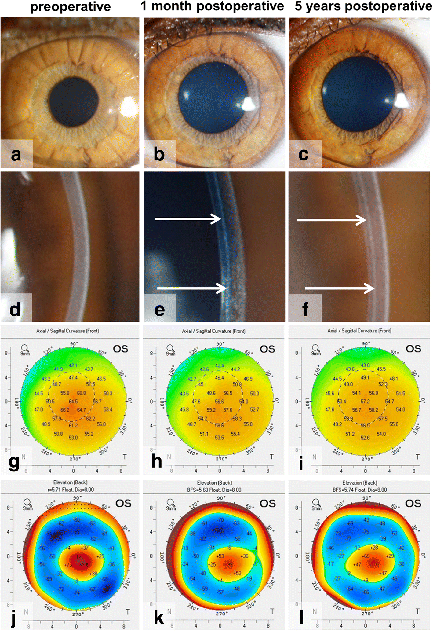
Bowman layer transplantation in the treatment of keratoconus | Eye and  Vision | Full Text
Bowman layer transplantation in the treatment of keratoconus | Eye and Vision | Full Text

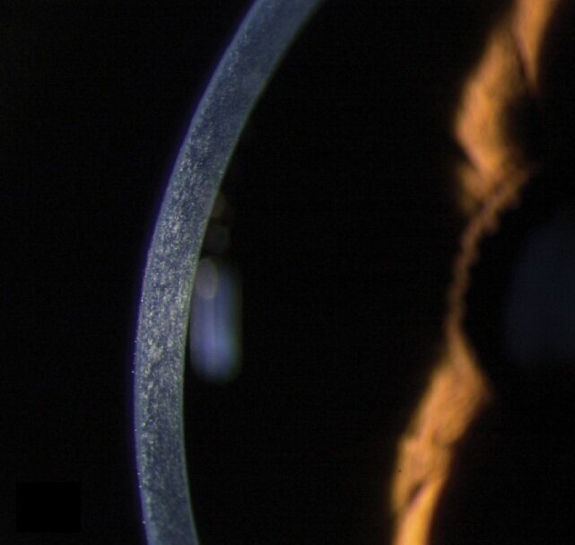
Cornea and anterior eye assessment with slit lamp biomicroscopy, specular  microscopy, confocal microscopy, and ultrasound biomicroscopy | Semantic  Scholar
Cornea and anterior eye assessment with slit lamp biomicroscopy, specular microscopy, confocal microscopy, and ultrasound biomicroscopy | Semantic Scholar

Top tips for slit-lamp imaging - part 2

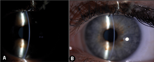
Figure, slit lamp image of cornea, iris and lens.] - StatPearls - NCBI  Bookshelf
Figure, slit lamp image of cornea, iris and lens.] - StatPearls - NCBI Bookshelf

Slit-lamp examination showed an irregular corneal surface and several... |  Download Scientific Diagram
Slit-lamp examination showed an irregular corneal surface and several... | Download Scientific Diagram

Optician Online - CPD Archive

The following is a slit lamp photograph of a 60 year old woman with  complaints of chronic foreign body sensation. Which of the following is the  least likely cause of her condition?
The following is a slit lamp photograph of a 60 year old woman with complaints of chronic foreign body sensation. Which of the following is the least likely cause of her condition?

Photographs of the cornea from six individuals examined using slit lamp...  | Download Scientific Diagram
Photographs of the cornea from six individuals examined using slit lamp... | Download Scientific Diagram
Slit-lamp Biomicroscopy

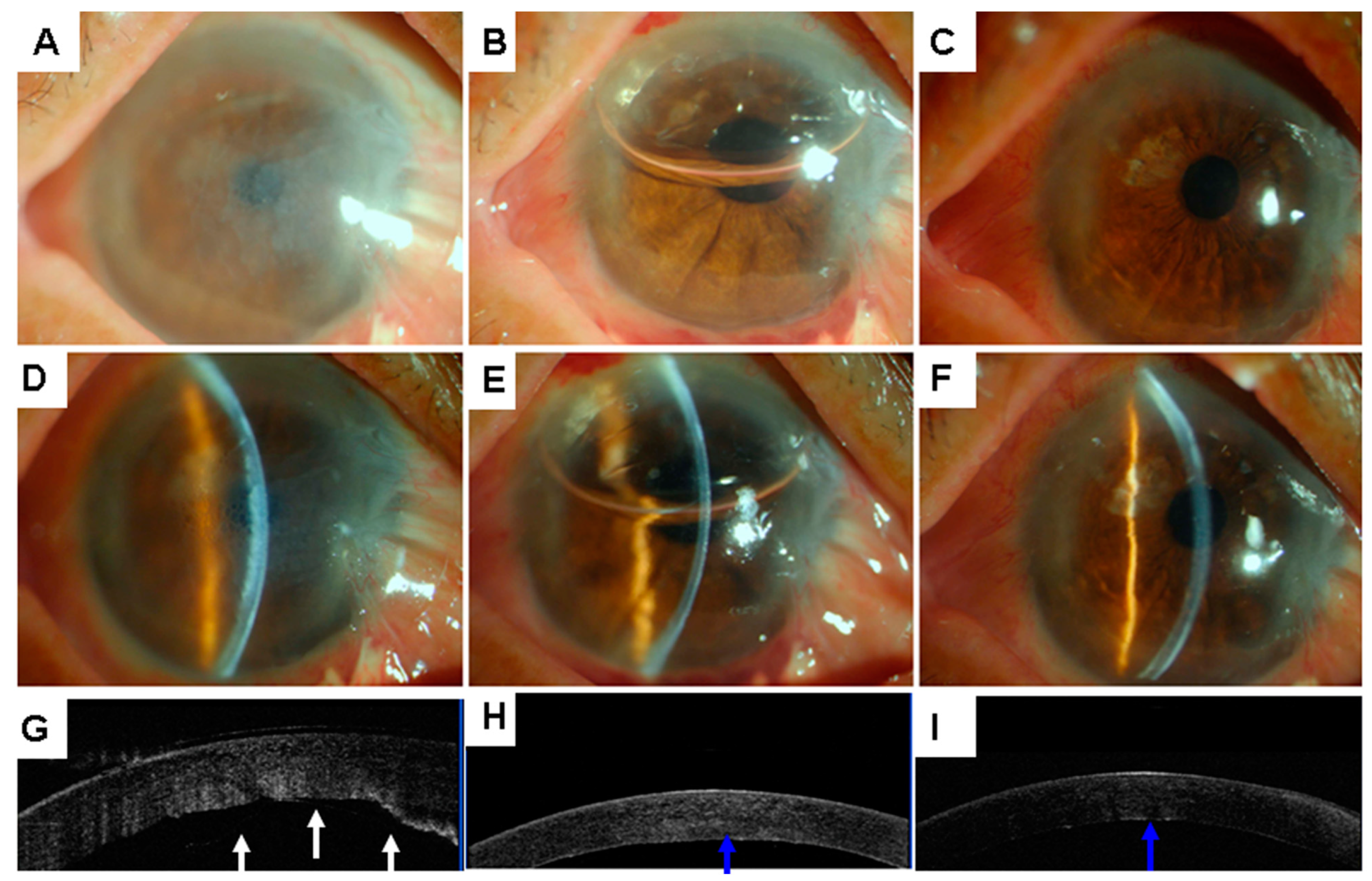
JCM | Free Full-Text | A Simple Repair Algorithm for Descemet’s  Membrane Detachment Performed at the Slit Lamp
JCM | Free Full-Text | A Simple Repair Algorithm for Descemet’s Membrane Detachment Performed at the Slit Lamp
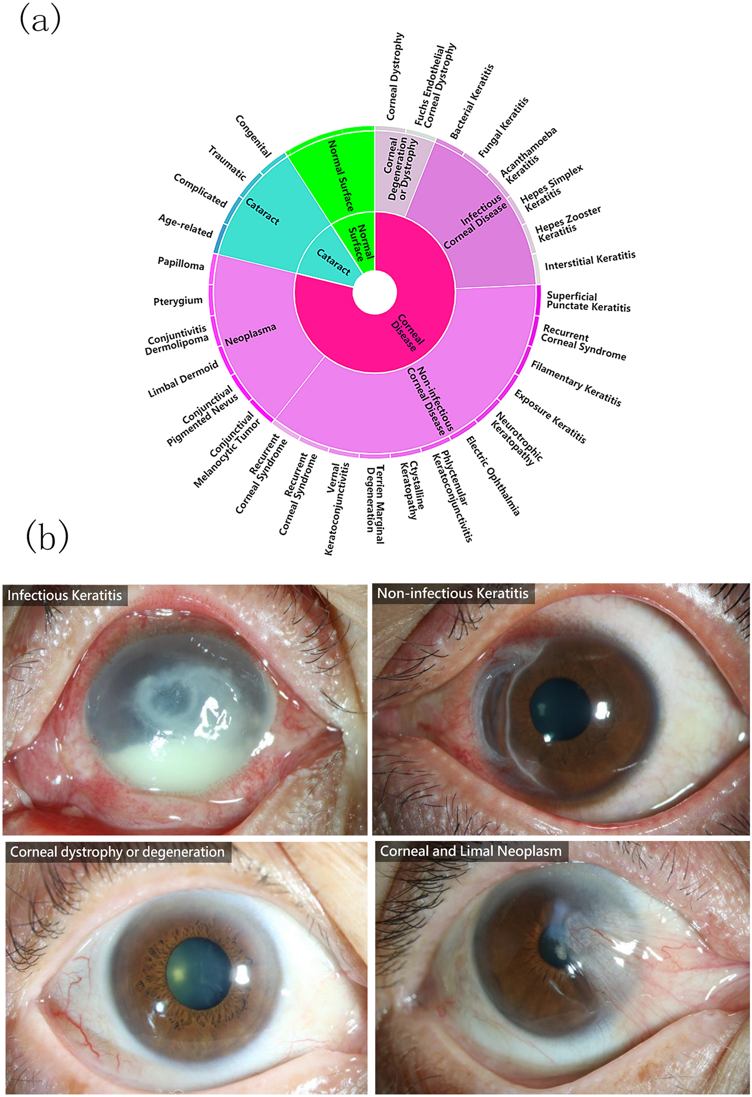
Deep learning for identifying corneal diseases from ocular surface slit-lamp  photographs | Scientific Reports
Deep learning for identifying corneal diseases from ocular surface slit-lamp photographs | Scientific Reports
Advanced Slit Lamp Skills

Ep 3: The Five (Possibly Six?) Layers of the Cornea — The Cornea Corner

Fine-Tune Your Corneal Disease Diagnostic Skills

Ophthalmology Dx: Blurred Sight for Sore Eye- Ophthalmology Advisor

Atlas Entry - Normal cornea

Slit-lamp exam Information | Mount Sinai - New York

Cornea slit lamp biomicroscopy cross-section art digital art

Slit lamp examination | Ento Key

Video slit lamp biomicroscope | Barnard Levit Optometrists

Lecture: Using the Slit Lamp Microscope to Visualize the Ocular Structures  - YouTube
Lecture: Using the Slit Lamp Microscope to Visualize the Ocular Structures - YouTube

Atlas Entry - Normal cornea

Fungal corneal ulcer (Video) - TimRoot.com

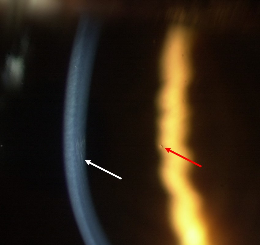
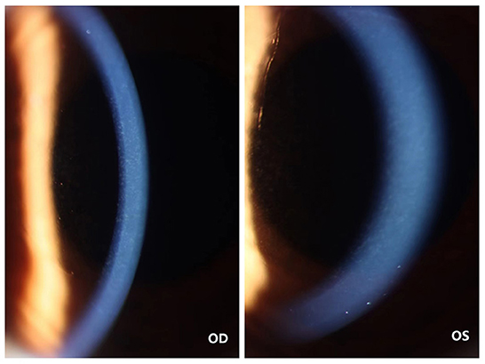
Frontiers | Case report: A case of corneal deposits between binocular  descemet membrane and corneal endothelial layer after small-incision  lenticule extraction (SMILE) followed by HPV vaccine
Frontiers | Case report: A case of corneal deposits between binocular descemet membrane and corneal endothelial layer after small-incision lenticule extraction (SMILE) followed by HPV vaccine